How is a Ureteral Stent Placed?

If you ever wondered how ureteral stents are placed, we’ll explain the process to you here step by step. Ureteral stents are typically used in two situations: 1) When a stone is causing blockage of the ureter and drainage needs to be established. 2) After a surgery to improve healing of the ureter or kidney.

How to place a ureteral stent in 8 steps (with video below):
- Use a cystoscope to locate the ureteral orifice where urine drains into the bladder. (A cystoscope is a camera that can be placed into the bladder).
- Pass a soft flexible wire called a “guidewire” into the ureteral orifice.
- Use x-ray imaging to monitor the guidewire as it is advanced up the ureter.
- If there is a blocking stone, contrast fluid can be injected using a soft hollow temporary stent. The contrast fluid will outline the ureter to improve guidance. The fluid can also push the stone back up the ureter in some cases.
- Advance the guidewire all the way into the kidney
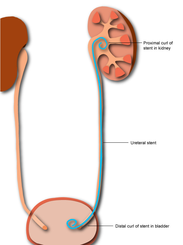
- Thread a ureteral stent over the guidewire and push it up into the kidney using a “pusher”.
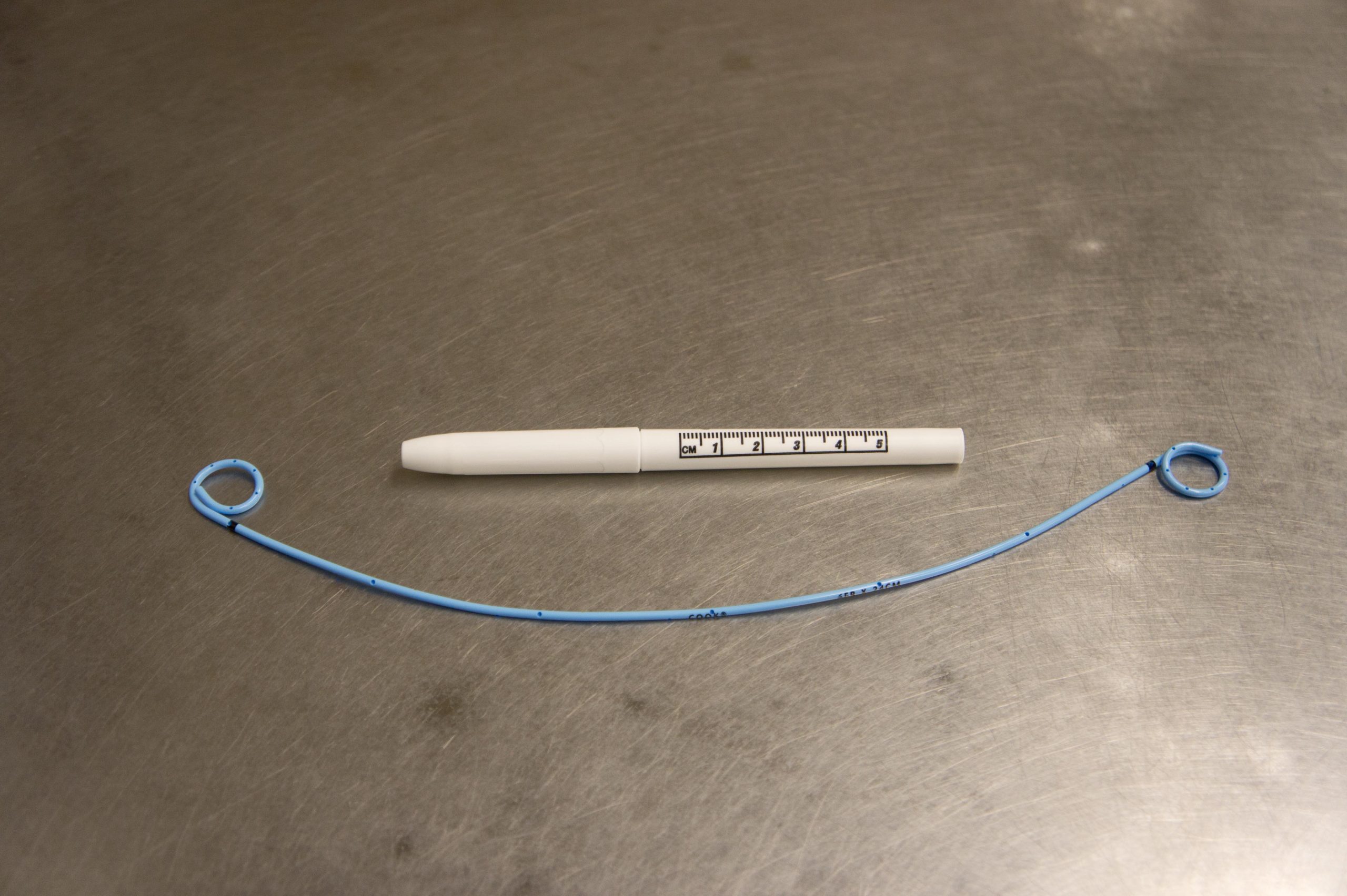
- Remove the guidewire once the ureteral stent is in the correct position. The stent has a natural curl at both ends which will keep it in place. Once the guidewire is removed, the curls can develop.
- Confirm correct placement of the stent using x-rays and by visualizing the stent in the bladder.
Video of ureteral stent placement in a patient with an obstructing left kidney stone in the ureter.

