
Kidney Stone Gallery
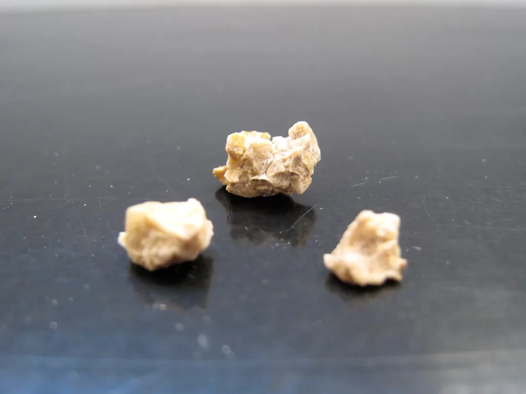 3 stone fragments that were removed during percutaneous surgery.
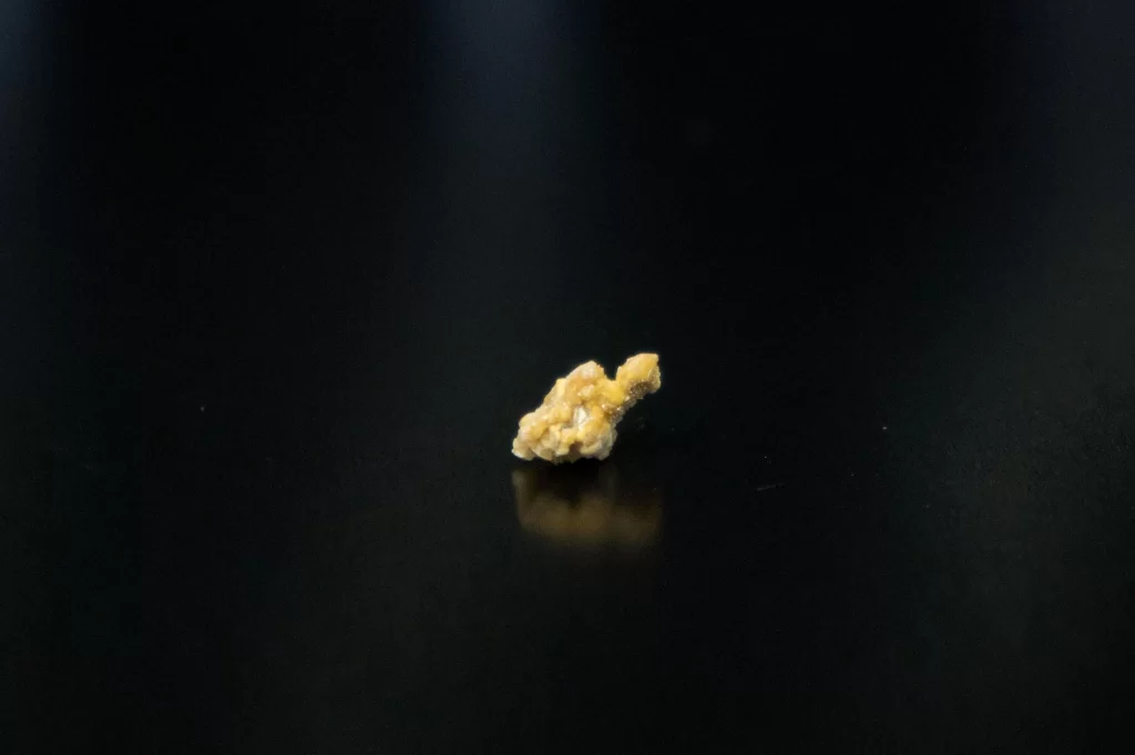 A 7 mm stone successfully passed by a patient. Comprised of calcium oxalate and calcium phosphate.
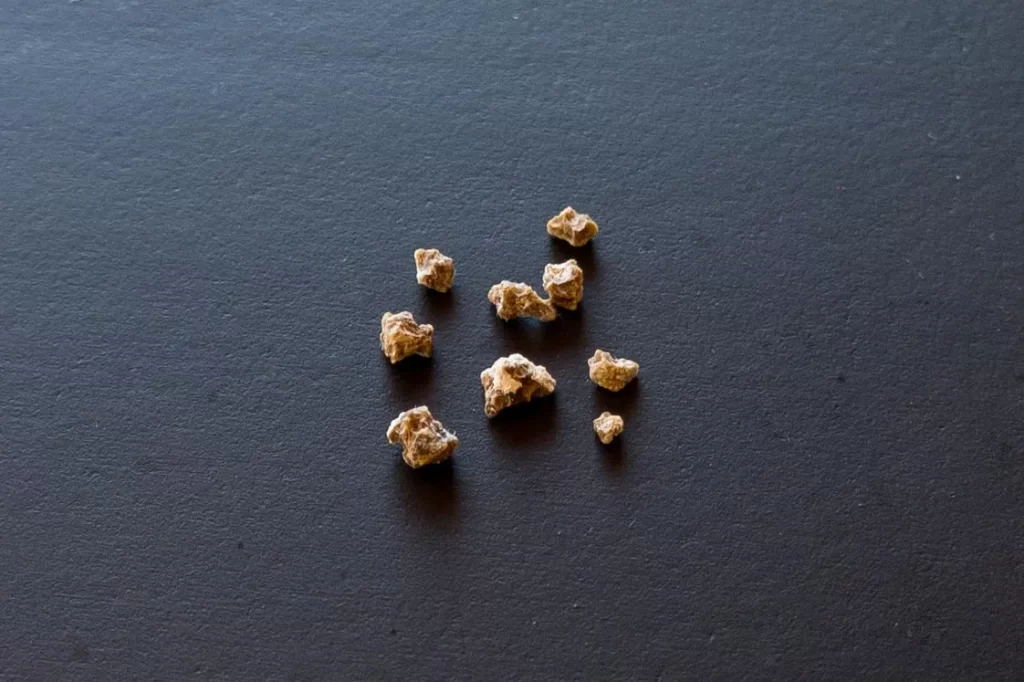 Fragments of an orginally 1.1 cm stone treated with laser lithotripsy and removed during ureteroscopy. Comprised of calcium oxalate.
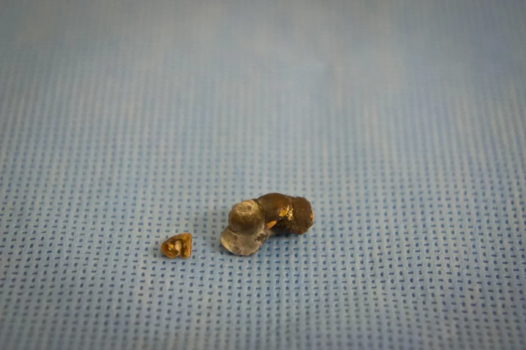 A 1.4 cm and a 5 mm stone removed percutaneously from a kidney.
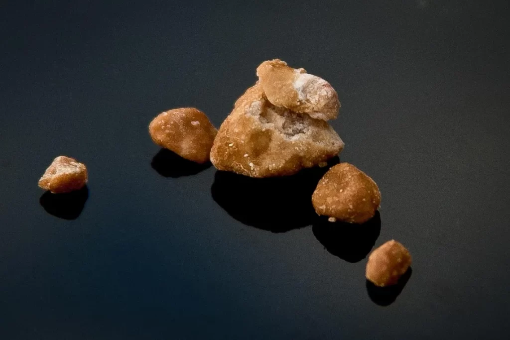 Collection of carbonate apatite stones removed percutaneously from a kidney.
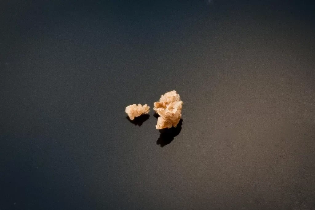 2mm and 4mm uric acid stones removed with ureteroscopy.
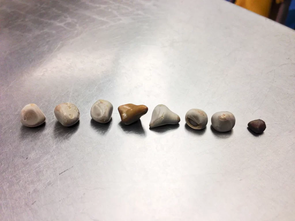 Multiple large smooth kidney stones removed percutaneously from a single kidney. Largest stone measures 1.2 cm in size. Analysis demonstrated 70% calcium oxalate monohydrate and 30% calcium phosphate.
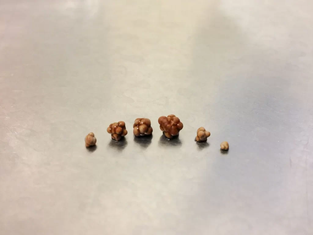 Multiple small kidney stones with the appearance of bird seed. Removed with ureteroscopy from a single kidney. Largest stone measured 4mm in size. Analysis demonstrated 85% calcium oxalate monohydrate and 15% calcium phosphate.
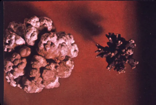 File photo of large bladder stones removed by open incision. These have a “jackstone” appearance.
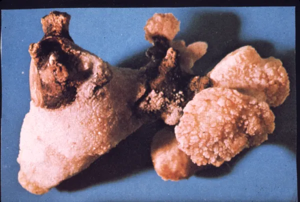 File photo of a large “staghorn” kidney stone removed intact by open surgery. The various parts of this stone fill up an entire kidney’s central collecting system, giving it a characteristic shape appearing similar to a deer’s antlers. Stones this size are not commonly seen intact anymore as most are now treated percutaneously and broken up before being removed.
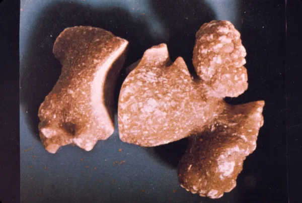 File photo of another large “staghorn” stone removed by open surgery.